The temporomandibular joint (TMJ) is a complex and highly functional joint responsible for enabling a wide range of mandibular movements such as chewing, speaking, and yawning. The intricate anatomy of the TMJ involves various components, including the articular disc, mandibular condyle, temporal bone, and several muscles, notably the lateral pterygoid muscle. Understanding the pathophysiology of TMJ disorders (TMD) requires insight into how these components interact, particularly when dysfunction occurs. Magnetic Resonance Imaging (MRI) has emerged as an indispensable tool for evaluating the internal structures of TMJ, especially in assessing articular disc displacement and the associated muscular changes. This article explores how MRI aids in the comprehensive assessment of articular disc displacement and the lateral pterygoid muscle, emphasizing recent findings and clinical implications.
Understanding Articular Disc Displacement
Definition and Significance
The articular disc in the TMJ is a fibrocartilaginous structure that divides the joint capsule into superior and inferior synovial spaces. Its primary functions include facilitating smooth movement of the mandibular condyle during jaw motions and absorbing mechanical load. Displacement of this disc—from its normal position—can lead to various clinical presentations, including joint pain, clicking sounds, limited mandibular movement, and, in severe cases, joint degeneration.
Types of Disc Displacement
- Simple anterior displacement: The most common form, where the disc is displaced anteriorly relative to the condyle.
- Displacement with reduction: The disc displaces anteriorly during mandibular opening but reduces back into its normal position during closing, often producing clicking sounds.
- Displacement without reduction: The disc remains displaced irreducibly, leading to restricted movement and potential joint issues.
Role of MRI in Evaluating TMJ Pathologies
Advantages of MRI
MRI provides superior soft tissue contrast, making it the gold standard for TMJ assessment. It allows for detailed visualization of the articular disc, condyle, joint capsule, and surrounding musculature without exposure to ionizing radiation. MRI sequences such as proton density and T2-weighted images are particularly effective in identifying disc position, joint effusion, and inflammatory changes.
Assessment of Articular Disc Displacement
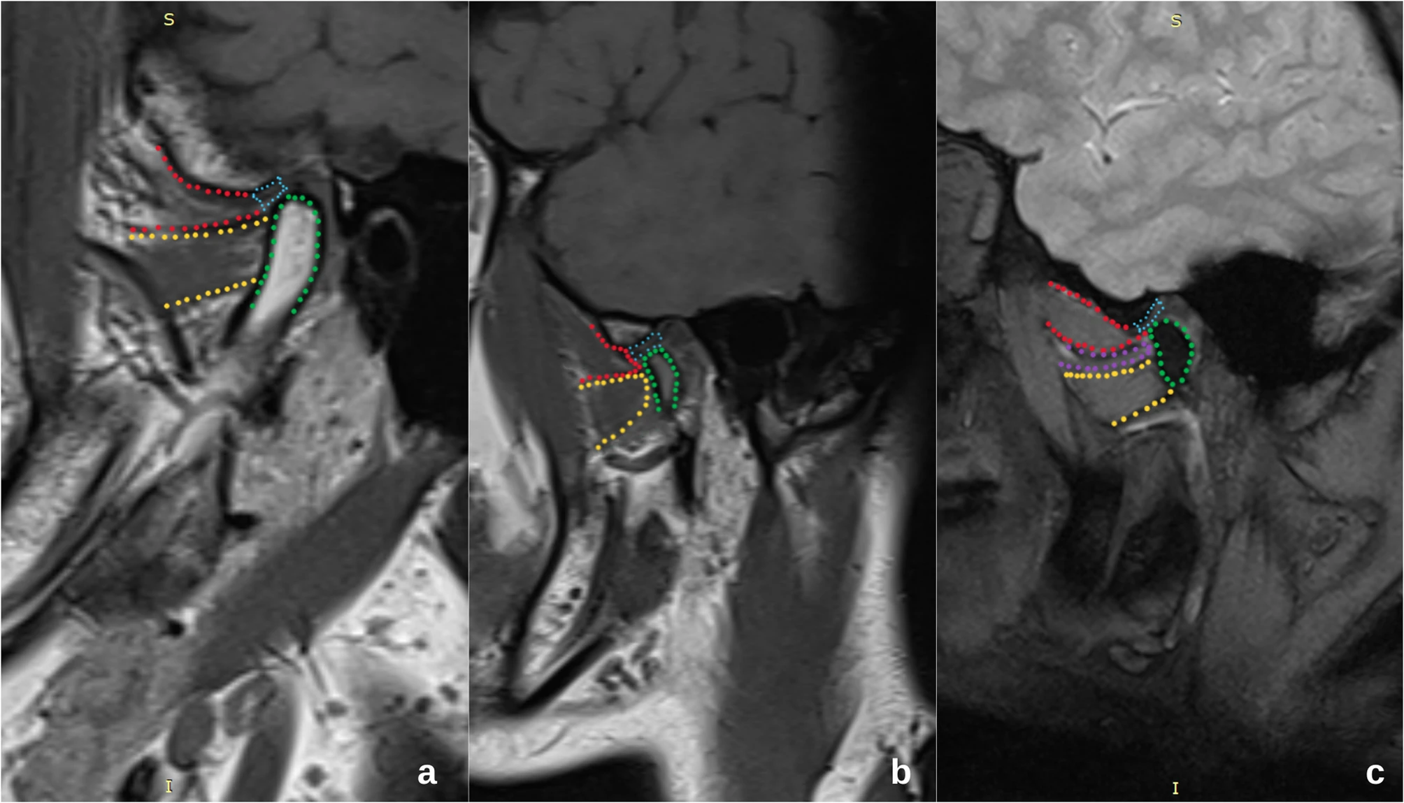
Using MRI, clinicians can categorize the precisely displaced position of the articular disc, determine whether it is reducible, and assess any associated morphological changes. The imaging typically involves both closed-mouth and open-mouth positions to evaluate dynamic movement of the disc during jaw motion.
Evaluating the Lateral Pterygoid Muscle in Relation to Disc Displacement
Importance of the Lateral Pterygoid Muscle
The lateral pterygoid muscle plays a critical role in mandibular movements, especially protrusion and lateral excursions. It also influences the position and stability of the articular disc. Dysfunction or abnormal activity of this muscle can contribute significantly to disc displacement and TMJ instability.
MRI Findings on the Lateral Pterygoid Muscle
MRI assessment extends beyond the disc itself to include the lateral pterygoid muscle. Changes such as muscle hypertrophy, atrophy, or abnormal signal intensity can indicate muscle overuse, fatigue, or inflammation, which may correlate with disc displacement patterns. Specifically, an **increased thickness or irregularities** may suggest compensatory mechanisms or muscular dysfunction contributing to joint instability.
Correlation Between Disc Displacement and the Lateral Pterygoid Muscle
Recent Research Findings
A notable study published in BMC Oral Health explored the relationship between articular disc displacement and the morphology and function of the lateral pterygoid muscle using MRI. The findings indicated that patients with disc displacement, particularly those without reduction, often exhibited significant alterations in the lateral pterygoid muscle, such as hypertrophy or abnormal signal intensity, compared to asymptomatic controls.
This correlation suggests that muscular factors may both influence and be affected by disc position. Abnormal activity of the lateral pterygoid may exert excessive anterior pull on the disc, leading to displacement. Conversely, chronic disc displacement may cause muscular adaptation, resulting in hypertrophy or fibrotic changes.
Clinical Implications
- Diagnosis & Treatment Planning: Understanding the relationship assists clinicians in devising targeted therapies focusing on muscular rehabilitation or behavioral modifications.
- Prognostic Value: Certain patterns of muscle changes may predict the likelihood of disc reduction or progression of TMJ dysfunction.
- Therapeutic Interventions: Muscle relaxation techniques, physiotherapy, or botulinum toxin injections may be employed to alleviate muscular hyperactivity contributing to disc displacement.
Integration of Imaging Findings into Clinical Practice
Comprehensive Approach
An integrated approach combining MRI findings of both the articular disc and lateral pterygoid muscle enhances diagnostic accuracy. It allows for differentiation between purely disc-related issues and muscular contributions, leading to personalized treatment strategies.
Future Directions
Ongoing research aims to develop standardized MRI protocols, quantitative assessments of muscle morphology, and dynamic imaging methods to better understand TMJ pathologies. Advances in functional MRI and 3D modeling hold promise for more precise diagnostics, predictive assessments, and minimally invasive interventions.
Conclusion
Magnetic resonance imaging stands at the forefront of TMJ disorder assessment, offering detailed insights into the complex interactions between the articular disc and lateral pterygoid muscle. Recognizing the correlation between disc displacement and muscular changes is vital for accurate diagnosis, effective treatment planning, and improved patient outcomes. A multidisciplinary approach integrating imaging findings with clinical evaluation remains essential for managing TMJ disorders comprehensively.
References: Evaluation of the correlation between articular disc displacement and the lateral pterygoid muscle using magnetic resonance imaging – Evaluation of the correlation between articular disc displacement and the lateral pterygoid muscle using magnetic resonance imaging BMC Oral Health
For more updated news please keep visiting Hourly Prime News.